Follicle After Ovulation Ultrasound
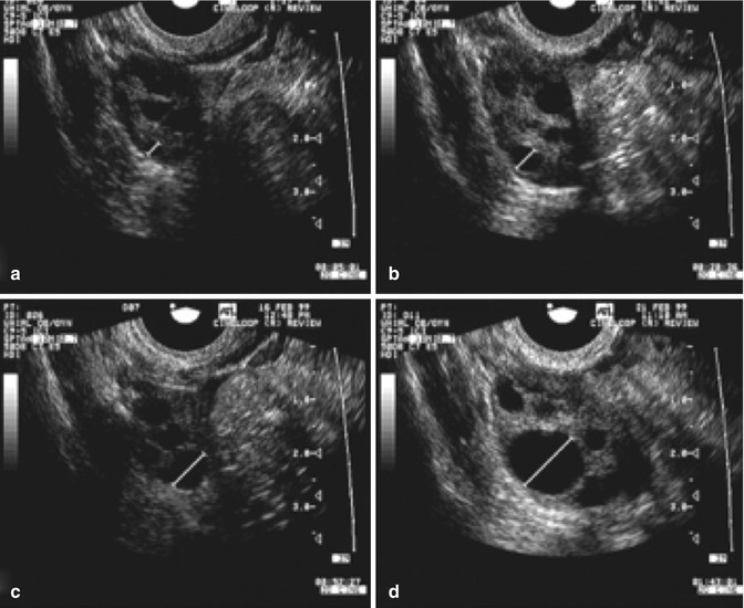
Follicle After Ovulation Ultrasound - Follicular tracking uses ultrasound to monitor the development of ovarian follicles during the first half of the menstrual cycle. In cases where a follicle doesn’t release an egg, it can result in a follicular cyst, which may be visible on an ultrasound. Next step is documentation of ovulation. Once the follicle reaches 16 mm size, a daily monitoring of follicle is recommended.
In cases where a follicle doesn’t release an egg, it can result in a follicular cyst, which may be visible on an ultrasound. Next step is documentation of ovulation. Once the follicle reaches 16 mm size, a daily monitoring of follicle is recommended. Follicular tracking uses ultrasound to monitor the development of ovarian follicles during the first half of the menstrual cycle.
Once the follicle reaches 16 mm size, a daily monitoring of follicle is recommended. Next step is documentation of ovulation. In cases where a follicle doesn’t release an egg, it can result in a follicular cyst, which may be visible on an ultrasound. Follicular tracking uses ultrasound to monitor the development of ovarian follicles during the first half of the menstrual cycle.

Ovarian Follicle Ultrasound
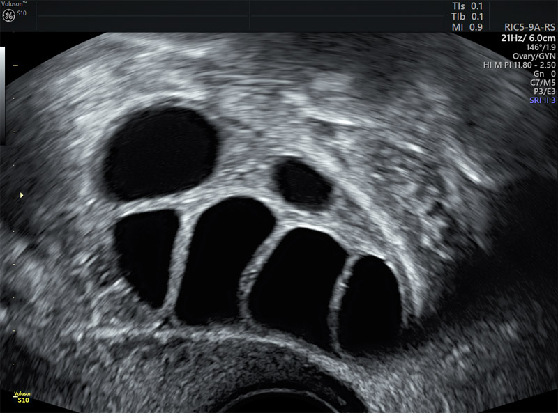
Follicular tracking uses ultrasound to monitor the development of ovarian follicles during the first half of the menstrual cycle. Once the follicle reaches 16 mm size, a daily monitoring of follicle is recommended. In cases where a follicle doesn’t release an egg, it can result in a follicular cyst, which may be visible on an ultrasound. Next step is documentation.

Overview of Follicular Scan For Ovulation Kiran IVF
Once the follicle reaches 16 mm size, a daily monitoring of follicle is recommended. Next step is documentation of ovulation. Follicular tracking uses ultrasound to monitor the development of ovarian follicles during the first half of the menstrual cycle. In cases where a follicle doesn’t release an egg, it can result in a follicular cyst, which may be visible on.
Ultrasound in Follicle Monitoring for Ovulation Induction/IUI
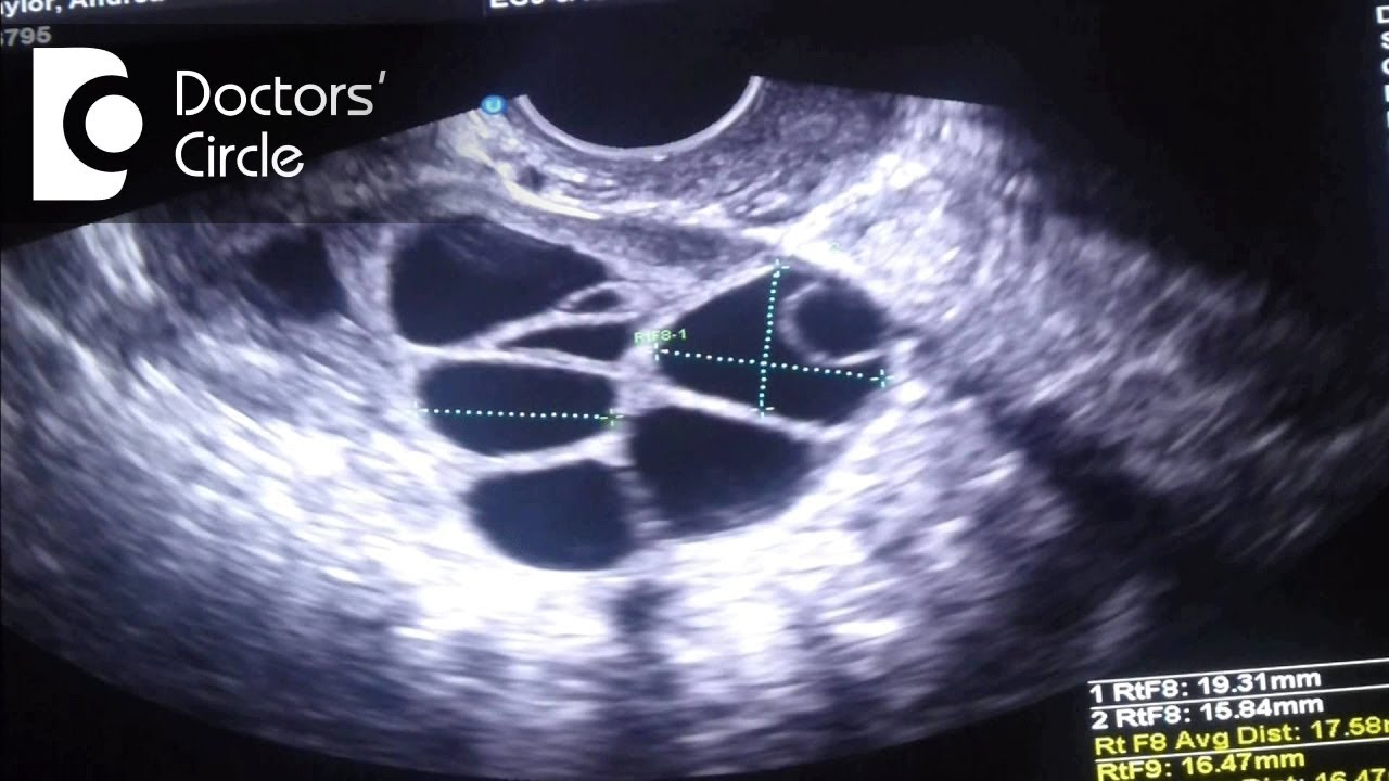
Follicular tracking uses ultrasound to monitor the development of ovarian follicles during the first half of the menstrual cycle. In cases where a follicle doesn’t release an egg, it can result in a follicular cyst, which may be visible on an ultrasound. Next step is documentation of ovulation. Once the follicle reaches 16 mm size, a daily monitoring of follicle.
Follicle Measurements Ultrasound Empowered Women's Health
Next step is documentation of ovulation. Follicular tracking uses ultrasound to monitor the development of ovarian follicles during the first half of the menstrual cycle. Once the follicle reaches 16 mm size, a daily monitoring of follicle is recommended. In cases where a follicle doesn’t release an egg, it can result in a follicular cyst, which may be visible on.
What is the ideal size of mature follicle for infertility treatment
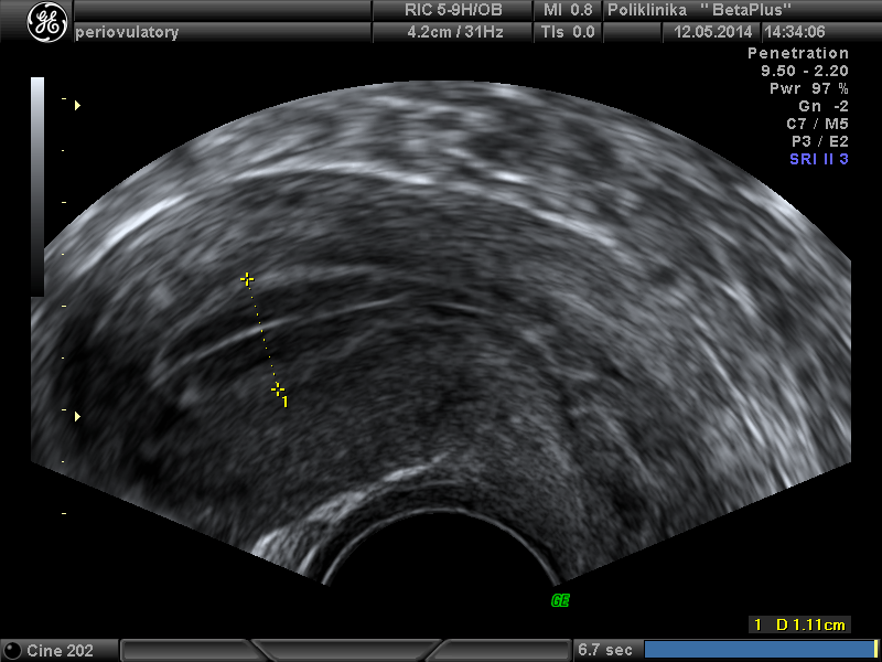
Follicular tracking uses ultrasound to monitor the development of ovarian follicles during the first half of the menstrual cycle. Once the follicle reaches 16 mm size, a daily monitoring of follicle is recommended. Next step is documentation of ovulation. In cases where a follicle doesn’t release an egg, it can result in a follicular cyst, which may be visible on.

TVS Ultrasound Follicular monitoring Raptured Follilce Ovulation
Next step is documentation of ovulation. Once the follicle reaches 16 mm size, a daily monitoring of follicle is recommended. In cases where a follicle doesn’t release an egg, it can result in a follicular cyst, which may be visible on an ultrasound. Follicular tracking uses ultrasound to monitor the development of ovarian follicles during the first half of the.

Signs Of Ovulation On Ultrasound
Once the follicle reaches 16 mm size, a daily monitoring of follicle is recommended. Follicular tracking uses ultrasound to monitor the development of ovarian follicles during the first half of the menstrual cycle. In cases where a follicle doesn’t release an egg, it can result in a follicular cyst, which may be visible on an ultrasound. Next step is documentation.

Automated follicle count using three‐dimensional ultrasound in
Once the follicle reaches 16 mm size, a daily monitoring of follicle is recommended. Next step is documentation of ovulation. Follicular tracking uses ultrasound to monitor the development of ovarian follicles during the first half of the menstrual cycle. In cases where a follicle doesn’t release an egg, it can result in a follicular cyst, which may be visible on.

Ultrasound scanning of ovulation Folliculometry (+ Photo
Next step is documentation of ovulation. Once the follicle reaches 16 mm size, a daily monitoring of follicle is recommended. Follicular tracking uses ultrasound to monitor the development of ovarian follicles during the first half of the menstrual cycle. In cases where a follicle doesn’t release an egg, it can result in a follicular cyst, which may be visible on.
US Follicle Monitoring BetaPlus Center for Reproductive Medicine
In cases where a follicle doesn’t release an egg, it can result in a follicular cyst, which may be visible on an ultrasound. Next step is documentation of ovulation. Follicular tracking uses ultrasound to monitor the development of ovarian follicles during the first half of the menstrual cycle. Once the follicle reaches 16 mm size, a daily monitoring of follicle.
Follicular Tracking Uses Ultrasound To Monitor The Development Of Ovarian Follicles During The First Half Of The Menstrual Cycle.
Once the follicle reaches 16 mm size, a daily monitoring of follicle is recommended. In cases where a follicle doesn’t release an egg, it can result in a follicular cyst, which may be visible on an ultrasound. Next step is documentation of ovulation.